| CPC A61B 8/5246 (2013.01) [A61B 8/0841 (2013.01); A61B 8/0883 (2013.01); A61B 8/12 (2013.01); A61B 8/4245 (2013.01); A61B 8/461 (2013.01); A61B 8/5284 (2013.01); A61B 8/543 (2013.01)] | 14 Claims |

|
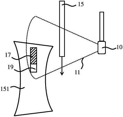
1. An ultrasound image processing apparatus comprising an image processor arrangement adapted to:
receive a plurality of ultrasound images obtained by an ultrasound probe, each ultrasound image imaging an invasive medical device relative to an anatomical feature of interest within a patient body during a particular phase of a cardiac cycle, said anatomical feature of interest having a shape that is different at different phases of the cardiac cycle, the plurality of ultrasound images covering at least two cardiac cycles during which the invasive medical device is displaced relative to the anatomical feature of interest and the ultrasound probe;
compile a plurality of groups of the ultrasound images, wherein the ultrasound images in each group belong to a same phase of said at least two cardiac cycles; and
for each group:
determine the displacement of the invasive medical device relative to the anatomical feature of interest between a first ultrasound image and a second ultrasound image in said group, wherein the shape of the anatomical feature of interest is the same in the first ultrasound image and the second ultrasound image and the ultrasound probe is stationary relative to the patient body such that a change between the first ultrasound image and the second ultrasound image is attributable to the displacement of the invasive medical device;
identify a first set of coordinates representative of an edge of an acoustic shadow region occurring within the first ultrasound image as a result of the displacement;
identify a corresponding second set of coordinates within the second ultrasound image;
determine whether the edge of the acoustic shadow region is present within the second ultrasound image at the second set of coordinates;
in response to determining that the edge of the acoustic shadow region is not present at the second set of coordinates, overlay one or more pixels occurring at the second set of coordinates from the second ultrasound image on the first ultrasound image at the first set of coordinates, thereby generating an augmented ultrasound image; and
output the augmented ultrasound image to a display.
|