| CPC A61B 5/341 (2021.01) [A61B 5/287 (2021.01); A61B 5/339 (2021.01); A61B 5/6858 (2013.01); A61B 5/6859 (2013.01)] | 20 Claims |

|
1. A method, comprising:
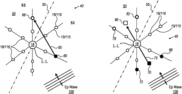
receiving (i) multiple electrophysiological (EP) signals acquired by multiple electrodes of a multi-electrode catheter that are in contact with tissue in a region of a cardiac chamber, and (ii) respective tissue locations at which the electrodes acquired the EP signals, the catheter including a longitudinal axis;
dividing the region into two sections with a virtual plane containing the longitudinal axis of the catheter;
using the EP signals acquired by the electrodes, calculating local activation time (LAT) values for the respective tissue locations, and finding a first section of the two sections having a smaller average LAT value, and a second section of the two sections having a higher average value;
determining a first representative location in the first section, and a second representative location in the second section;
calculating between the first and second representative locations a propagation vector indicative of propagation of an EP wave that has generated the EP signals; and
presenting the propagation vector to a user in a graphical form.
|