| CPC A61B 6/5217 (2013.01) [A61B 5/02007 (2013.01); A61B 5/08 (2013.01); A61B 6/032 (2013.01); A61B 6/481 (2013.01); A61B 6/504 (2013.01); A61B 6/507 (2013.01); A61B 6/508 (2013.01); A61B 6/541 (2013.01); G06T 7/0012 (2013.01); G06T 7/0016 (2013.01); G06T 7/11 (2017.01); G06T 7/143 (2017.01); G06T 7/187 (2017.01); G06T 7/35 (2017.01); G06T 7/62 (2017.01); A61B 5/026 (2013.01); A61B 5/113 (2013.01); A61B 5/7285 (2013.01); A61B 2576/02 (2013.01); G06T 2207/10076 (2013.01); G06T 2207/10081 (2013.01); G06T 2207/10088 (2013.01); G06T 2207/10136 (2013.01); G06T 2207/20016 (2013.01); G06T 2207/20024 (2013.01); G06T 2207/20076 (2013.01); G06T 2207/30061 (2013.01); G06T 2207/30101 (2013.01); G06T 2207/30104 (2013.01); G06T 2207/30172 (2013.01)] | 14 Claims |
|
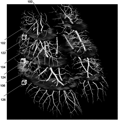
1. A method of calculating a ventilation/perfusion ratio from at least one in vivo lung image comprising a vasculature tree and acquired in the absence of contrast agent and a time series of lung images, the method comprising:
applying a filter to the at least one in vivo lung image comprising the vasculature tree to provide a probability field and a scale field;
performing vessel segmentation on the probability field to extract a segmented vasculature tree from the probability field;
mapping the scale field to the segmented vasculature tree to quantify a geometry of the vasculature tree;
measuring a motion of a portion of the lung from the time series of lung images; and
comparing the motion of the portion of the lung to the geometry of the vasculature tree in the region of the portion of the lung to obtain the ventilation/perfusion ratio.
|