| CPC A61B 5/444 (2013.01) [A61B 5/0035 (2013.01); A61B 5/0075 (2013.01); A61B 5/443 (2013.01); A61B 5/7264 (2013.01); A61B 8/0858 (2013.01)] | 1 Claim |

|
1. A method, comprising the following steps:
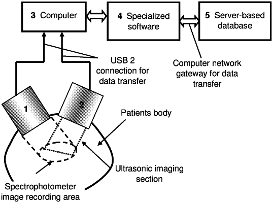
recording first images, comprising dermatoscopic and individual skin chromophore spatial distribution images, by using a spectrophotometric device to emit light of different wavelengths onto an area of interest of skin;
recording second images and data, comprising B-mode images and data of the area of interest of the skin, using an ultrasonic imaging device operating at a frequency over 20 MHz for imaging structures beneath a surface of the skin;
compiling a database, including the first images, the second images and data, and histological data for classifying skin tumors;
classifying the area of interest of the skin using a computer processor that is configured to:
load the first images recorded by the spectrophotometric device and the second images and data recorded by the ultrasonic imaging device,
distinguish a tumor area in the first images recorded by the spectrophotometric device, wherein a blue component of the first images recorded by the spectrophotometric device is used to define the tumor area,
determine a depth of the tumor area by the second images and data recorded by the ultrasonic imaging device,
estimate quantitative parameters of the first images recorded by the spectrophotometric device, including each of parameterizing the first images and evaluating parameters of a surface shape of the tumor area, wherein spectral parameters of the tumor area, tumor form parameters, and image texture parameters of a first range and a second range of internal sections of the tumor area are used to parameterize the second images and data recorded by the ultrasonic imaging device, and
use the estimated quantitative parameters and the database including the first images, the second images and data, and the histological data for classifying skin tumors, to classify and report the tumor area within the area of interest of the skin as one of a malignant tumor or a non-malignant tumor.
|