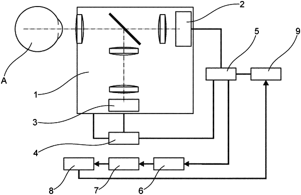
| CPC A61B 3/12 (2013.01) [A61B 3/0008 (2013.01); A61B 3/14 (2013.01); A61B 3/1241 (2013.01)] | 7 Claims |

|
1. A method for examining a retinal vascular endothelial function of vessels of a retina at a fundus (F) of an eye (A), the method comprising:
adjusting a fundus camera to the eye (A);
generating measuring light for illuminating the vessels of the retina at the fundus (F) and generating illuminating flicker light for stimulation of the vessels of the retina at the fundus (F) during a stimulation phase (SP);
generating an image sequence of images of an area of the fundus (F) during a baseline phase (BP), at least one stimulation phase (SP) and at least one posterior phase (NP);
measuring vessel diameters of selected vascular segments of the vessels of the retina in the images of the image sequence as a function of location and time;
performing movement correction for the vascular segments, wherein each vascular segment is assigned to a location on the fundus (F) in a movement-corrected manner;
forming diameter signals (D(t,x,y)) representing the measured vessel diameters as a function of the time and location of each selected vascular segment; and
deriving as vascular parameters the maximum flicker dilation from the diameter signals (D(t,x,y)) of each selected vascular segment, each of said vascular parameters describing the endothelial function of a respectively selected vascular segment;
wherein illuminating and stimulating a macula (M) at the fundus (F) is prevented by projecting a sharp image of one macula stop (MB) with a fixation mark (FM) attached to it onto the fundus (F), and aligning the eye (A) with said one macula stop (MB) by fixation on the fixation mark (FM) of said one macula stop (MB) such that the macula (M) is covered by an image of said one macula stop (MB).
|