| CPC G01N 33/6842 (2013.01) [G01N 33/6848 (2013.01); G16B 45/00 (2019.02); G01N 2333/4716 (2013.01); G01N 2570/00 (2013.01)] | 39 Claims |
|
1. A method of proteome analysis comprising the steps of:
obtaining a biological sample, wherein the biological sample is an animal tissue sample, animal biopsy, animal cell homogenate, animal cell fraction, cultured animal cells, non-cultured animal cells, animal whole blood, animal plasma, animal biological fluids, or a single cell organism;
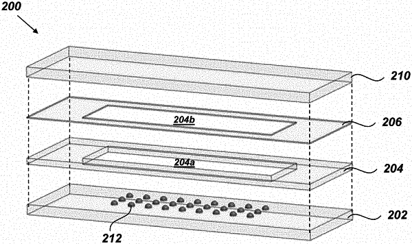
transferring a first volume of the biological sample to a platform, wherein the platform comprises a substrate comprising at least one reactor vessel having one or more hydrophilic surfaces configured for containment of a biological sample, wherein the hydrophilic surfaces have a non-zero total surface area less than 25 mm2, a spacer plate containing a spacer plate aperture, wherein the spacer plate aperture is dimensioned to surround the at least one reactor vessel when the spacer plate is positioned on the substrate, a cover positioned on the spacer plate, and a membrane interposed between the spacer plate and the cover and containing a membrane aperture that is dimensioned to surround the at least one reactor vessel when the membrane is positioned on the spacer plate, wherein the substrate, spacer plate and cover are each formed from a material that is transparent to optical light and wherein transferring the first volume of the biological sample comprises transferring the first volume of the biological sample to the at least one reactor vessel, wherein the first volume is a non-zero amount less than 1000 nL;
processing the biological sample in the at least one reactor vessel;
extracting from the at least one reactor vessel a processed sample comprising less than 500 ng of a complement of proteins, peptides related to the complement of proteins, or both;
dispensing the processed sample into one well on a well plate having a plurality of wells, wherein the one well is pre-loaded with a volume of a liquid carrier buffer and the volume of the processed sample that is transferred into one well on the well plate is a non-zero amount that is less than 50 μL;
diluting the processed sample, thereby yielding in the one well a diluted sample, wherein the diluted sample has a volume of at least 5 μL, and the diluting comprises dispensing a volume of a wash solution into the at least one reactor vessel and subsequently transferring the at least one reactor vessel's contents to the one well;
transferring the diluted sample from the one well to a mass-spectrometry-based analytical instrument; and
providing protein identification for each of a plurality of proteins composing the complement of proteins.
|