| CPC B23P 19/047 (2013.01) [A61B 17/1214 (2013.01); A61B 17/12031 (2013.01); A61B 17/12113 (2013.01); A61B 17/12145 (2013.01); A61B 17/12172 (2013.01); A61B 17/12186 (2013.01); A61B 17/12168 (2013.01); A61B 2017/00526 (2013.01); A61B 2017/00557 (2013.01); A61B 2017/00862 (2013.01); A61B 2017/00929 (2013.01); A61B 2017/12054 (2013.01); A61B 2017/12063 (2013.01); A61B 2090/376 (2016.02); A61B 2090/3966 (2016.02)] | 17 Claims |
|
1. A method for treatment of an aneurysm, the method comprising:
acquiring a first image visualizing:
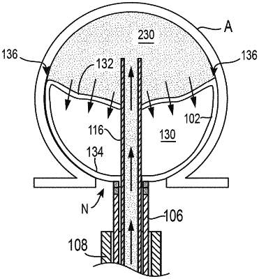
an occlusive member positioned within the aneurysm, the occlusive member including a first radiopaque marker and substantially filling a volume of the aneurysm; and
a conduit having a distal portion positioned within the aneurysm, the distal portion of the conduit including a second radiopaque marker; and
deforming the occlusive member so that the occlusive member collapses on itself by delivering an embolic element between the occlusive member and a dome of the aneurysm via the conduit; and
acquiring a second image in which the first radiopaque marker is further from the second radiopaque marker than in the first image, indicating collapse of the occlusive member.
|