| CPC A61B 8/12 (2013.01) [A61B 5/6852 (2013.01); A61B 8/4483 (2013.01); A61B 8/461 (2013.01); A61M 25/0084 (2013.01); A61M 25/0097 (2013.01); A61M 25/01 (2013.01); A61M 25/0662 (2013.01); A61M 39/10 (2013.01)] | 20 Claims |
|
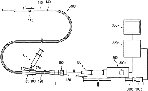
1. A method of using a diagnostic imaging catheter, the diagnostic imaging catheter comprising: a sheath having a lumen and a proximal end;
an axially movable drive shaft positioned in the lumen of the sheath and possessing a distal portion at which is located a transducer unit, the drive shaft also possessing an outer circumference and a proximal end; a relay connector coupled to the proximal end of the sheath; a support tube covering the outer circumference of at least a portion of an axial extent of the drive shaft, the support tube being movable forward and backward together with axial movement of the drive shaft; an injection opening communicating with the lumen in the sheath; a port in communication with the injection opening; a unit connector positioned proximal of the relay connector; an outer tube extending between the relay connector and the unit connector, the drive shaft passing through the outer tube, at least a part of the support tube being positioned inside the outer tube; and a hub positioned proximal of the relay connector and connected to the drive shaft to rotate the drive shaft and axially move the drive shaft forward and backward; the method comprising:
pulling the hub in a proximal direction to a maximum extent and thereby also axially moving the drive shaft and the transducer unit in the proximal direction, the pulling of the hub in the proximal direction causing the drive shaft to axially move relative to the outer tube;
connecting a syringe containing fluid to the port, the syringe including a plunger;
pushing the plunger of the syringe to inject the fluid into the relay connector via the injection opening, the fluid which is injected into the relay connector flowing into the sheath;
discharging the fluid outside of the sheath via a discharge hole in the sheath;
connecting a connector portion of the diagnostic imaging catheter to an external apparatus;
pushing the hub in a distal direction to a maximum extent and thereby also axially moving the drive shaft and the transducer unit in the distal direction, the pushing of the hub in the distal direction causing the drive shaft to axially move relative to the outer tube;
positioning the sheath at a target position inside a body cavity of a living body;
acquiring a tomographic image inside the body cavity of the living body by moving the transducer unit in the proximal direction in a pullback operation while also rotating the transducer unit; and
the support tube covering the outer circumference of at least the portion of the axial extent of the drive shaft such that the support tube covers the drive shaft at the injection opening during at least a part of the pullback operation to prevent the drive shaft from entering the injection opening.
|