| CPC A61B 3/0025 (2013.01) [A61B 3/107 (2013.01); G06T 2207/10101 (2013.01)] | 5 Claims |
|
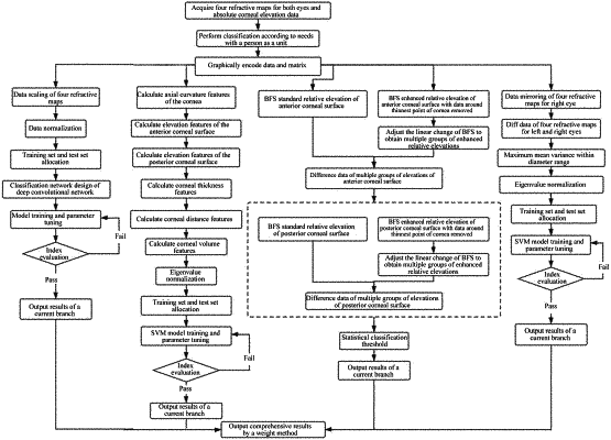
1. A method for early diagnosis of keratoconus based on multi-modal data, specifically comprising the following steps:
1) acquiring binocular multi-modal data comprising four refractive maps for corneas of both eyes and absolute corneal elevation data, wherein the four refractive maps for the corneas comprise an axial curvature map of an anterior corneal surface, a relative elevation topography of the anterior corneal surface, a relative elevation topography of a posterior corneal surface, and a corneal thickness topography; and the absolute corneal elevation data comprises absolute elevation data of the anterior and posterior corneal surfaces;
2) according to classified cases, associating the binocular multi-modal data with classification categories, and classifying the data according to requirements;
3) unifying data of topographies and the elevation data of the anterior and posterior corneal surfaces in the four refractive maps in step 2) into data matrices of a same size;
4) based on the above data, determining early keratoconus of the both eyes using four branch methods, which are respectively recorded as a branch A method, a branch B method, a branch C method, and a branch D method; wherein
the branch A method is as follows: after data processing, sending all of the data matrices of the four refractive maps to a classification network of a deep convolutional network to identify sensitivity and specificity of the keratoconus and obtain a classification result P(A) output for a certain case;
the branch B method is as follows: calculating eigenvalues of each graphic data matrix in all of the data matrices of the four refractive maps, and sending eigenvalue data to a binary classification method using a support vector machine (SVM) to identify the sensitivity and specificity of the keratoconus and obtain a classification result P(B) output for the certain case;
the branch C method is as follows: comparing the absolute elevation data of the anterior and posterior corneal surfaces with best-fit-sphere (BFS) data to obtain a critical threshold between keratoconus cases and normal cases, so as to determine a classification result P(C) output for the certain case; and
the branch D method is as follows: obtaining optimal sensitivity and specificity as well as a probability P(D) of the keratoconus of the certain case using the critical threshold or using an SVM classification method by taking an average value, a maximum value, and a standard deviation of the data matrices of the four refractive maps for left and right eyes as feature quantities; and
5) weighting and accumulating final results in the branch A method, the branch B method, the branch C method and the branch D method to obtain a final probability of the keratoconus of the certain case.
|