| CPC G06T 7/0016 (2013.01) [G06V 10/62 (2022.01); G06V 10/895 (2022.01); G06T 2207/30104 (2013.01); G06V 2201/07 (2022.01)] | 20 Claims |

|
1. A method of imaging a target, comprising:
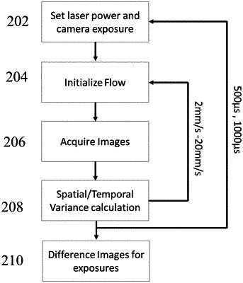
acquiring, by a processor of an imaging apparatus, multiple images of the target, wherein the multiple images have different exposure values;
determining temporal variances for the multiple images, wherein the temporal variances are determined over pixel regions having predetermined dimensions over a pre-defined number of images over a period of time, and wherein the temporal variance of a given pixel region represents a difference between a square of mean square values of the pixels in the given pixel region over a pre-defined number of frames, N, where N is a positive integer, and a mean value of square values of the pixels over the pre-defined number of frames;
determining spatial variances for the multiple images, wherein the spatial variances are determined over the pixel regions having predetermined dimensions, and wherein a spatial variance of a given pixel region represents a difference between a mean value of squares of pixel values and a square of a mean value of pixels in the region; and
generating a perfusion image of the target by combining the temporal variances and the spatial variances such that a local flow rate in the perfusion image at a given pixel is a function of changes in the spatial variances and the temporal variances as a function of exposure values;
wherein the generating the perfusion image comprises evaluating an average according to:
 wherein N is a total number of exposures collected, IPERF represents pixel value at each pixel location of the perfusion image, IEiSPAT/TEMP and IEi+iSPAT/TEMP represent spatial or temporal variances at corresponding pixel location for exposure times Ei and Ei+i, WEi and WEi+i are real numbers representing a relative weight for each exposure.
|