| CPC A61B 6/5205 (2013.01) [A61B 5/0044 (2013.01); A61B 5/349 (2021.01); A61B 6/032 (2013.01); A61B 6/461 (2013.01); A61B 6/481 (2013.01); A61B 6/503 (2013.01); A61B 6/504 (2013.01); A61B 6/5264 (2013.01); G06T 7/0014 (2013.01); G06T 7/74 (2017.01); G16H 30/20 (2018.01); G16H 30/40 (2018.01); G06T 2207/20081 (2013.01); G06T 2207/30048 (2013.01)] | 39 Claims |

|
1. A method for generating an image of an object of interest of a patient, the object of interest comprising a heart, a part of the coronary tree, blood vessels or other part of the vasculature of the patient, the method comprising:
i) obtaining first image data of the object of interest, wherein the first image data is acquired using an X-ray imaging modality with a contrast agent and an interventional device is present in the first image data, the interventional device being used in a procedure to treat the object of interest, wherein the first image data covers at least one cardiac cycle of the patient;
ii) using the first image data to generate a plurality of roadmaps of the object of interest;
iii) determining a plurality of reference locations of a tip of the interventional device in the first image data, wherein the plurality of reference locations correspond to the plurality of roadmaps of the object of interest;
iv) obtaining second image data of the object of interest, wherein the second image data is acquired using an X-ray imaging modality without a contrast agent and the interventional device is present in the second image data;
v) selecting a roadmap from the plurality of roadmaps;
vi) determining a location of the tip of the interventional device in the second image data;
vii) using the reference location of the tip of the interventional device corresponding to the roadmap selected in v) and the location of the tip of the interventional device determined in vi) to transform the roadmap selected in v) to generate a dynamic roadmap of the object of interest; and
viii) overlaying a visual representation of the dynamic roadmap of the object of interest as generated in vii) on the second image data for display;
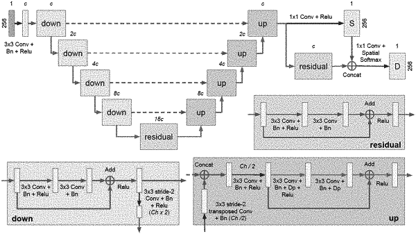
wherein, in vi), the location of the tip of the interventional device in the second image data is determined by inputting the second image data to a trained machine learning network that outputs a posterior probability distribution that estimates likelihood of the location of the tip of the interventional device in the second image data given the second image data as input and using a Bayesian filtering method that equates location of the tip of the interventional device to a weighted arithmetic mean of a plurality of positions and their associated weights derived from the posterior probability distribution output by the trained machine learning network.
|