| CPC G06T 7/0012 (2013.01) [G06T 2207/10101 (2013.01); G06T 2207/30041 (2013.01); G06T 2207/30101 (2013.01)] | 12 Claims |
|
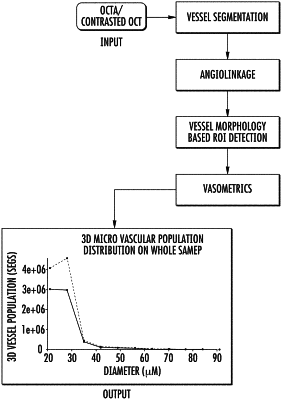
1. A method of identifying a 3D region of interest of a retina from a plurality of retinal blood vessels in a 3D optical coherence tomography (OCT) image, comprising more than one OCT image, of at least a portion of the retina, the method comprising:
using at least one computer programmed to perform:
obtaining a geometrical representation of a vascular tree or vessel network of retinal blood vessels from the 3D OCT image;
evaluating at least one first morphological feature of a plurality of retinal blood vessels detected in the geometrical representation of the 3D OCT image at each of a plurality of voxel locations in the geometrical representation; and
identifying a first region of interest based at least in part on evaluating the at least one first morphological feature of the plurality of retinal blood vessels, wherein the first region of interest comprises a particular retinal layer,
wherein evaluating at least one first morphological feature comprises determining a feature field comprising the at least one morphological feature at each of a plurality of voxel locations, and wherein identifying the first region of interest comprises evaluating the feature field.
|