| CPC A61B 8/5207 (2013.01) [A61B 8/02 (2013.01); A61B 8/06 (2013.01); A61B 8/4236 (2013.01); A61B 8/4254 (2013.01); A61B 8/5223 (2013.01); A61B 8/5276 (2013.01)] | 27 Claims |

|
1. A system comprising:
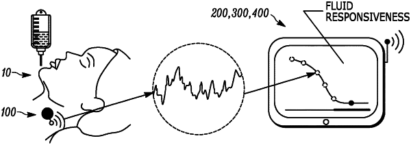
a patch-type imaging ultrasound sensor configured to attach to a patient;
an ultrasound scanning system including a single-element transducer configured to acquire low-level ultrasound data from the sensor such that at least one of 2 dimensional planes or 3-dimensional volumes are automatically acquired; and
a processing system configured to perform a data acquisition sequence in which the low-level ultrasound data is collected and to perform signal and image processing of the low-level ultrasound data to automatically convert the low level ultrasound data into a numerical measurement;
wherein the sensor is communicatively coupled to the ultrasound scanning system and the processing system, the processing system configured to utilize a swarm speckle tracking approach to:
automatically measure tissue motion of a tissue to determine a presence or absence and respective location of at least one vein,
discriminate between other vessels and identify a presence or absence of a specific vein within a pre-specified anatomic region,
identify a vessel wall region of the specific vein,
automatically select a plurality of tracking markers residing at locations near the vessel wall region,
automatically assign a unique pseudorandom thrust vector to each of the plurality of tracking markers,
automatically track and analyze a movement of the tracking markers over at least one respiratory and cardiac cycle, wherein the movement is representative of temporal geometric changes, and
compute a contractility index based on the temporal geometric changes of the vessel wall region, wherein the contractility index is computed by computing a ratio between:
(i) a difference between a maximum geometry and a minimum geometry of the vessel wall region, and
(ii) the maximum geometry,
over the at least one respiratory and cardiac cycle according to the movement of the tracking markers such that at least one measurement of the specific vein can be computed for each 2-dimensional plane or 3-dimensional volume obtained.
|