| CPC G01N 15/1463 (2013.01) [G01N 33/54306 (2013.01); G01N 2015/008 (2013.01); G01N 2015/0038 (2013.01); G01N 2015/1006 (2013.01); G01N 2015/1486 (2013.01); G01N 2333/4737 (2013.01)] | 48 Claims |

|
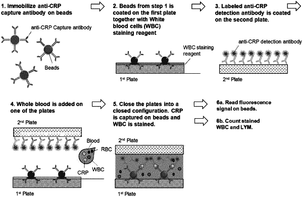
1. A method for assaying a targeted cell and a non-cell analyte in a sample, comprising:
(a) obtaining two plates facing each other and separated by a gap, wherein each of the plates has a sample contact area for contacting a sample;
(b) obtaining a cell staining reagent, a capture agent, and a sample that contains or is suspected of containing a targeted cell and a non-cell analyte;
(c) confining the cell staining reagent, and the capture agent, and the sample between the sample contact areas of the two plates, forming a thin sample layer, wherein, the sample has a thickness confined by the gap, wherein the cell staining reagent stains the targeted cell, and the capture agent specifically captures the non-cell analyte; and
(d) imaging, after (c), the thin sample layer to (i) detect and count the targeted cell that is stained by the cell staining reagent, and (ii) detect the non-cell analyte that is captured by the capture agent;
wherein the gap has a size configured to make the targeted cell have a monolayer in the thin sample layer and wherein, in the monolayer, the targeted cells do not have significant overlap.
|