| CPC H01J 37/28 (2013.01) [H01J 2237/2602 (2013.01); H01J 2237/28 (2013.01)] | 27 Claims |
|
1. A method for analyzing a specimen in a microscope, the method comprising:
acquiring a series of compound image frames using an electron detector and a second detector, the second detector comprising any of an X-ray spectrometer, an electron diffraction pattern camera, an electron energy loss spectrometer, or a cathodoluminescence detector, wherein acquiring a compound image frame comprises:
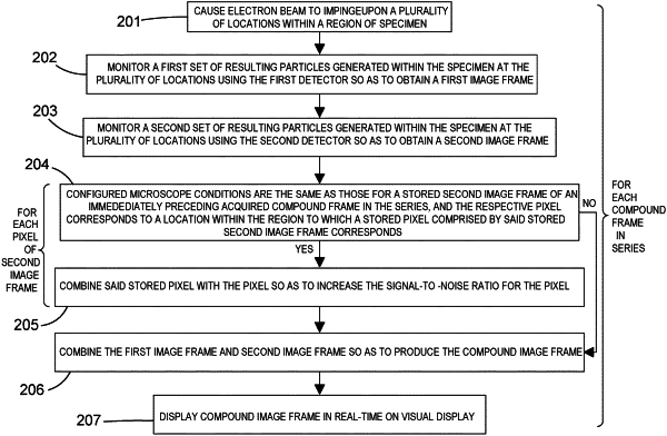
a) causing a charged particle beam to impinge upon a plurality of locations within a region of a specimen, the region corresponding to a configured field of view of the microscope, the microscope being configured with a set of microscope conditions,
b) monitoring, in accordance with the configured microscope conditions, a first set of resulting particles generated within the specimen at the plurality of locations using the electron detector so as to obtain a first image frame,
c) monitoring, in accordance with the configured microscope conditions, a second set of resulting particles generated within the specimen at the plurality of locations using the second detector, so as to obtain a second image frame, wherein each image frame comprises a plurality of pixels corresponding to, and having values derived from the monitored particles generated at, the plurality of locations within the region,
d) for each pixel of the second image frame:
when the configured microscope field of view is different from that for an immediately preceding compound image frame in the series, using the value of the pixel to replace the value for a corresponding pixel in a stored second image frame; and
when the configured microscope field of view is the same as that for the immediately preceding compound image frame in the series, combining the value of the pixel with the value of the corresponding pixel in the stored second image frame so as to increase the signal-to-noise ratio for the corresponding pixel of the stored second image frame, and
e) combining the first image frame and stored second image frame so as to produce the compound image frame, such that the compound image frame provides data derived from, for each of the plurality of pixels, the particles generated at the corresponding location within the region and monitored by each of the electron detector and the second detector;
and displaying the series of compound image frames in real-time on a visual display,
wherein steps (b) and (c) are performed substantially simultaneously; and
wherein the visual display is updated to show each compound image frame in sequence so as to allow an observer to identify potential features of interest when the field of view is changing.
|
|
19. A system according to claim 17, wherein the short time period is less than 1 second.
|