| CPC A61B 6/5217 (2013.01) [G06T 7/0012 (2013.01); G06T 7/11 (2017.01); G16H 30/40 (2018.01); G16H 50/20 (2018.01); A61B 5/055 (2013.01); A61B 6/032 (2013.01); G06T 2207/10081 (2013.01); G06T 2207/10088 (2013.01); G06T 2207/20081 (2013.01); G06T 2207/20132 (2013.01); G06T 2207/30096 (2013.01)] | 26 Claims |
|
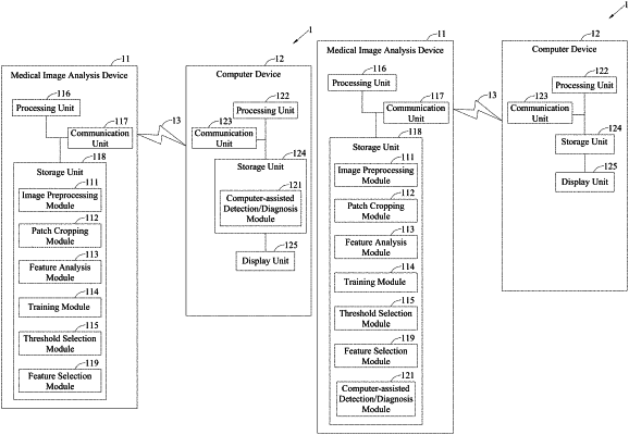
1. A medical image analyzing system, comprising:
an image preprocessing module, executed by a processor, configured to process at least one image corresponding to an organ to generate at least one processed image, wherein the processed image comprises a segmentation label corresponding to a cancerous part of the organ;
a patch cropping module, executed by the processor, configured to acquire a plurality of image patches from the processed image;
a feature analysis module, executed by the processor, configured to perform feature analysis on the plurality of image patches to obtain a plurality of feature values corresponding to each of the plurality of image patches;
a training module, executed by the processor, configured to train a full model using the plurality of feature values of each of the plurality of image patches to obtain a plurality of first prediction values corresponding to the respective plurality of image patches;
a threshold selection module, executed by the processor, configured to plot a first curve based on the plurality of first prediction values and determine a first threshold that is used for determining whether each of the plurality of image patches is cancerous according to the first curve; and
a computer-assisted detection/diagnosis module, executed by the processor, configured to input at least one patient image to the image preprocessing module and the patch cropping module to generate a plurality of patient image patches and input the plurality of patient image patches into the full model to obtain a plurality of first prediction values corresponding to the respective plurality of patient image patches,
wherein the computer-assisted detection/diagnosis module further enables the threshold selection module to calculate at least one second prediction value corresponding to the at least one patient image based on the plurality of first prediction values respectively corresponding to the plurality of patient image patches, and plot a second curve based on the at least one second prediction value to determine a second threshold that is used for determining whether the at least one patient image is cancerous according to the second curve.
|