| CPC A61B 8/085 (2013.01) [A61B 8/06 (2013.01); A61B 8/0891 (2013.01); A61B 8/4254 (2013.01); A61B 8/4444 (2013.01); A61B 8/4494 (2013.01); A61B 8/461 (2013.01); A61B 8/469 (2013.01); A61B 8/488 (2013.01); A61B 8/0858 (2013.01); A61B 2562/0219 (2013.01)] | 18 Claims |

|
1. An ultrasound imaging system, comprising:
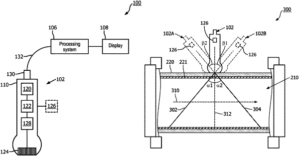
an imaging probe configured for handheld operation by a user;
an ultrasound transducer array within the imaging probe and configured to obtain imaging data associated with blood flow through a blood vessel within a body of a subject, wherein the imaging data comprises Doppler data;
a first display device on the imaging probe and comprising an indicator light; and
a processor within the imaging probe and in communication with the ultrasound transducer array and the first display device, wherein the processor configured to:
during a first portion of an imaging procedure:
receive the imaging data from the ultrasound transducer array while the imaging probe is moved to be in a plurality of orientations;
identify, based on the imaging data, a transition between a positive velocity of the blood flow and a negative velocity of the blood flow; and
determine, based on the transition, a perpendicular orientation of the imaging probe in which the imaging probe is positioned at a perpendicular angle with respect to the blood vessel; and
during a second portion of the imaging procedure that occurs after the first portion:
determine, using the perpendicular orientation as a reference, an angle of the imaging probe with respect to the blood vessel while the imaging probe is in a first orientation; and
output, to the first display device, a visual representation of the angle for the user, wherein the visual representation of the angle comprises a color of the indicator light, wherein the color is representative of a numerical value of the angle such that the indicator light changes to a different color when the imaging probe is moved to be in a second orientation in which the imaging probe is positioned at a different angle with respect to the blood vessel.
|