| CPC G06T 7/0012 (2013.01) [A61N 5/1031 (2013.01); G06T 5/002 (2013.01); G06T 7/11 (2017.01); G06T 2207/10081 (2013.01); G06T 2207/20021 (2013.01); G06T 2207/20182 (2013.01); G06T 2207/30064 (2013.01); G06T 2207/30168 (2013.01)] | 14 Claims |
|
1. A system for assessing a pulmonary image, the system comprising:
a memory that stores a plurality of instructions; and
at least one processor that couples to the memory and is configured to execute the plurality of instructions to:
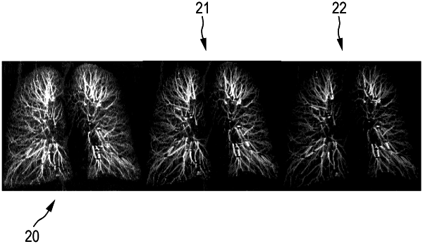
provide the pulmonary image comprising image elements having assigned image values, the pulmonary image showing lung vessels;
smooth the provided pulmonary image for providing different pulmonary images with different degrees of smoothing;
determine signal values for the different pulmonary images, wherein for a respective pulmonary image one or several signal values, which are indicative of detectability of the lung vessels in the respective pulmonary image, are determined based on the image values of the respective pulmonary image;
determine noise values for the different pulmonary images, wherein for the respective pulmonary image one or several noise values, which are indicative of the noise in the respective pulmonary image, are determined based on the image values of the respective pulmonary image;
determine an image quality for an unsmoothed pulmonary image based on the signal values and noise values determined for the different pulmonary images; and
determine a radiation dose level to be applied for generating a next pulmonary image based on the determined image quality.
|