| CPC G06T 7/0012 (2013.01) [A61B 6/502 (2013.01); A61B 6/5217 (2013.01); G06T 3/0093 (2013.01); G06T 2207/20081 (2013.01); G06T 2207/20084 (2013.01); G06T 2207/30068 (2013.01); G06T 2207/30096 (2013.01)] | 20 Claims |
|
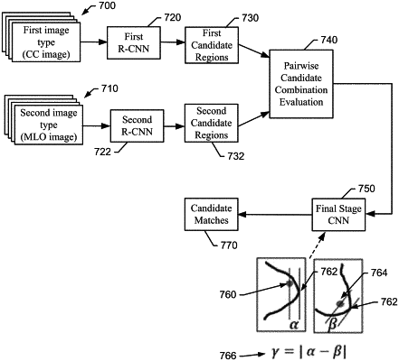
1. A method of identifying potential lesions in mammographic images, the method comprising:
receiving, by an image processing device, first image data;
receiving, by the image processing device, second image data, one of the first image data or the second image data being two-dimensional Craniocaudal (CC) mammographic image data or two-dimensional Mediolateral Oblique (MLO) mammographic image data;
registering, by the image processing device, the first image data and the second image data by employing an image registration convolutional neural network (CNN) using pixel level registration; wherein registering the first image data with the second image data comprises:
inputting the first image data and the second image data into the image registration CNN;
generating, via convolutions performed by the image registration CNN on the first image data and the second image data, a deformation field of deformation vectors that map pixels of the first image data to pixels of the second image data; the deformation field comprising, to define the deformation vectors,
a vertical deformation data array that defines row-wise relationships between the pixels of the first image data and the pixels of the second image data and
a horizontal deformation data array that defines column-wise relationships between the pixels of the first image data and the pixels of the second image data;
determining, by the image processing device, whether a candidate detection of a lesion exists in both the first image data and the second image data based on the first image data and the second image data and a mapping of the first image data to the second image data provided by deformation field output from the image registration CNN; and
generating, by the image processing device, display output identifying the lesion.
|