| CPC A61B 8/463 (2013.01) [A61B 8/06 (2013.01); A61B 8/145 (2013.01); A61B 8/488 (2013.01); A61B 8/5246 (2013.01); A61B 8/469 (2013.01); A61B 8/5223 (2013.01)] | 34 Claims |
|
1. A method for ultrasound flow imaging display, comprising:
transmitting focused ultrasound beams to a scanning target selected by a user;
receiving echoes of the focused ultrasound beams to obtain focused ultrasound echo signals;
obtaining ultrasound images of at least a portion of the scanning target according to the focused ultrasound echo signals;
displaying the ultrasound images on a display device;
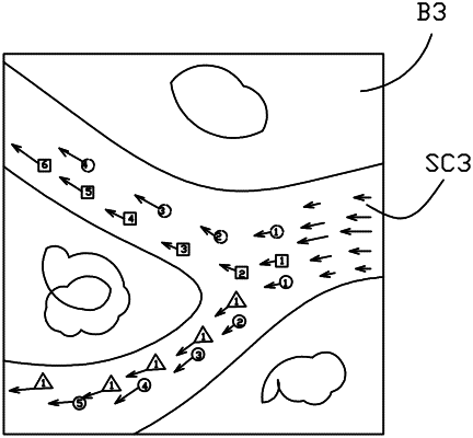
receiving, from the user, location information for a plurality of individual, user-selected target points within the user-selected scanning target, the user-selected target points comprising discrete, user-selected locations of moving particles of a blood flow within the user-selected scanning target;
transmitting plane ultrasound beams to the user-selected scanning target;
receiving echoes of the plane ultrasound beams to obtain plane ultrasound echo signals;
obtaining flow velocity vector information for the user-selected target points in the user-selected scanning target according to the plane ultrasound echo signals;
superimposing the ultrasound images and the flow velocity vector information to form vector flow images comprising a flow velocity vector for at least two moving particles selected by at least two user-selected target points, each of the at least two moving particles being graphically represented by a different fixed shape for uniquely distinguishing the at least two moving particles from each other over a time interval within the blood flow, wherein each flow velocity vector comprises an arrow, a length of the arrow representing a magnitude of the flow velocity vector of the respective moving particle and a direction of the arrow representing the direction of the flow velocity vector of the respective moving particle; and
displaying the vector flow images on the display device.
|