| CPC A61B 8/0891 (2013.01) [A61B 8/14 (2013.01); A61B 8/4209 (2013.01); A61B 8/4245 (2013.01); A61B 8/466 (2013.01); A61B 8/467 (2013.01); A61B 8/54 (2013.01); G06T 7/10 (2017.01); G06T 7/62 (2017.01); A61B 2562/0247 (2013.01); G06T 2207/10136 (2013.01); G06T 2207/30101 (2013.01); G06T 2207/30172 (2013.01)] | 20 Claims |

|
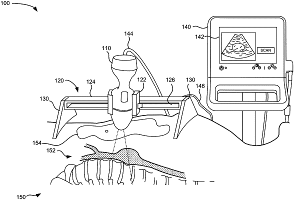
18. A non-transitory computer-readable medium having stored thereon sequences of instructions which, when executed by at least one processor, cause the at least one processor to:
communicate with an ultrasound probe to detect an installed orientation of the ultrasound probe secured with respect to a track structure and confirm that a gyroscopic reading of the ultrasound probe is consistent with the installed rotational orientation of the ultrasound probe installed in a probe holder of the track structure;
detect movement of the probe holder at increments along the track structure;
cause the ultrasound probe to transmit ultrasound signals at each of the increments while in contact with a patient;
record a linear position of the probe holder at each of the increments;
receive echo information associated with the transmitted ultrasound signals; and
process the received echo information to generate a three-dimensional ultrasound image of an extended target based on the received echo information and the linear positions of the probe holder,
wherein processing the received echo information includes:
obtaining a three-dimensional model corresponding to the extended target, wherein the three-dimensional model includes a statistical shape model that infers a statistical distribution and is derived from human samples,
identifying a best-fit of the three-dimensional model for the three-dimensional ultrasound image by minimizing an energy function,
storing the best fit of the three-dimensional model as a segmentation result, and
calculating, based on the segmentation result, a longitudinal centerline for the extended target.
|