| CPC G16H 30/40 (2018.01) [A61B 1/000094 (2022.02); A61B 1/000096 (2022.02); A61B 1/045 (2013.01); A61B 1/05 (2013.01); G06T 7/0012 (2013.01); G06T 2207/10068 (2013.01)] | 18 Claims |
|
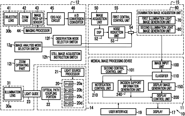
1. A medical image processing device comprising:
a processor configured to:
acquire an examination image of a subject captured by an endoscope;
identify an incision suitable site in the subject included in the examination image;
perform control for outputting incision suitable site information regarding the incision suitable site on the basis of the examination image;
generate, on the basis of the incision suitable site information, a first display image indicating the incision suitable site with a color, a symbol, or a figure;
superimpose the first display image on the examination image; and
output the first display image superimposed on the examination image to a display, wherein
the identification of the incision suitable site information is performed by using a learning image, the learning image being associated with information regarding a position of a muscular layer in the subject, the information regarding the position of the muscular layer in the subject including three-dimensional information indicating a thickness of the submucosal layer covering the surface of the muscular layer and two-dimensional information indicating a range of the muscular layer region in the learning image.
|