| CPC G06T 7/0012 (2013.01) [A61B 3/0025 (2013.01); A61B 3/0041 (2013.01); A61B 3/102 (2013.01); A61B 3/1225 (2013.01); G06T 3/20 (2013.01); G06T 2207/10101 (2013.01); G06T 2207/30041 (2013.01)] | 18 Claims |

|
1. An ophthalmologic information processing apparatus for analyzing an image of a subject's eye formed by arranging a plurality of A-scan images acquired by performing an Optical Coherence Tomography (OCT) scan on an inside the subject's eye with measurement light deflected around a scan center position, the ophthalmologic information processing apparatus comprising processing circuitry configured to:
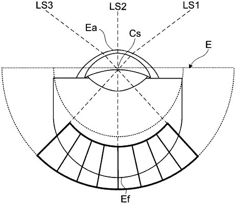
transform pixel positions in the image into transformation positions along traveling directions of the measurement light passing through the scan center position by transforming pixel positions in each A-scan image into transformation positions along the traveling directions of the measurement light used to form the A-scan image;
specify a layer region by analyzing the image in which the pixel position has been transformed; and
specify a normal direction of the layer region.
|