US 11,941,179 B2
Imaging system and method for use in surgical and interventional medical procedures
Robert E. Isaacs, San Diego, CA (US); Samuel Morris Johnston, San Diego, CA (US); David Alexander Skwerer, San Diego, CA (US); and Randall Graham Campbell, San Diego, CA (US)
Assigned to NuVasive, Inc., San Diego, CA (US)
Filed by NuVasive, Inc., San Diego, CA (US)
Filed on Dec. 13, 2021, as Appl. No. 17/549,059.
Application 17/549,059 is a continuation of application No. 16/857,069, filed on Apr. 23, 2020, granted, now 11,231,787.
Application 16/857,069 is a continuation of application No. 16/558,155, filed on Sep. 1, 2019, granted, now 10,684,697, issued on Jun. 16, 2020.
Application 16/558,155 is a continuation of application No. 16/200,519, filed on Nov. 26, 2018, granted, now 10,444,855, issued on Oct. 15, 2019.
Application 16/200,519 is a continuation of application No. 15/713,265, filed on Sep. 22, 2017, granted, now 10,139,920, issued on Nov. 27, 2018.
Application 15/713,265 is a continuation of application No. 14/564,728, filed on Dec. 9, 2014, granted, now 9,785,246, issued on Oct. 10, 2017.
Application 14/564,728 is a continuation in part of application No. 14/270,446, filed on May 6, 2014, granted, now 8,908,952, issued on Dec. 9, 2014.
Application 14/270,446 is a continuation of application No. 13/722,259, filed on Dec. 20, 2012, granted, now 8,718,346, issued on May 6, 2014.
Application 13/722,259 is a continuation in part of application No. 13/253,838, filed on Oct. 5, 2011, granted, now 8,526,700, issued on Sep. 3, 2013.
Claims priority of provisional application 62/036,660, filed on Aug. 13, 2014.
Claims priority of provisional application 61/390,488, filed on Oct. 6, 2010.
Prior Publication US 2022/0113810 A1, Apr. 14, 2022
This patent is subject to a terminal disclaimer.
Int. Cl. G06K 9/00 (2022.01); A61B 5/05 (2021.01); A61B 6/00 (2006.01); A61B 6/06 (2006.01); A61B 6/12 (2006.01); G06F 3/01 (2006.01); G06T 3/20 (2006.01); G06T 7/00 (2017.01); G06T 7/33 (2017.01); G06T 11/60 (2006.01); G06T 15/08 (2011.01); H04N 7/18 (2006.01)
CPC G06F 3/017 (2013.01) [A61B 6/06 (2013.01); A61B 6/12 (2013.01); A61B 6/4405 (2013.01); A61B 6/4441 (2013.01); A61B 6/486 (2013.01); A61B 6/5241 (2013.01); A61B 6/547 (2013.01); G06T 3/20 (2013.01); G06T 3/4053 (2013.01); G06T 7/0016 (2013.01); G06T 7/33 (2017.01); G06T 11/60 (2013.01); G06T 15/08 (2013.01); H04N 7/18 (2013.01); G06T 2207/10124 (2013.01); G06T 2207/20212 (2013.01); G06T 2207/20221 (2013.01); G06T 2207/30004 (2013.01); G06T 2210/41 (2013.01)] 18 Claims
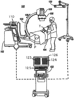
OG exemplary drawing
 
1. A method for generating a display of an image of a patient's internal anatomy in a surgical field during a medical procedure, the method comprising:
with one or more processors:
producing an image set using a machine, wherein the image set includes permutations of a first image, the first image being based on a three-dimensional representation of the surgical field including the patient's internal anatomy and being with respect to a baseline orientation;
applying a first dose of radiation to the patient to obtain the three-dimensional representation;
acquiring a new image of the surgical field by applying a second dose of radiation to the patient, wherein the first dose of radiation is greater than the second dose of radiation;
selecting a representative image from the image set based on a correlation between the representative image and the new image;
identifying, within the new image, a location of a glyph caused by a radiodense object coupled to an imaging device used to obtain the new image;
merging the selected representative image with the new image using the location of the glyph as part of the merging; and
displaying the merged image.