| CPC H04N 23/74 (2023.01) [A61B 46/10 (2016.02); A61B 90/30 (2016.02); H04N 23/51 (2023.01); H04N 23/56 (2023.01); H04N 23/71 (2023.01); A61B 2017/00477 (2013.01); A61B 2090/306 (2016.02); A61B 2090/309 (2016.02); A61B 90/361 (2016.02); A61B 2090/3941 (2016.02)] | 15 Claims |
|
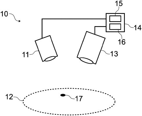
1. A method of displaying fluorescence intensity for imaged anatomy, the method comprising:
displaying images of the anatomy captured by an optical imaging device being aimed at the anatomy;
displaying a target reticle on the displayed images around a region of a field of view of the imaging device;
calculating a representation of normalized fluorescence intensity within the target reticle based on normalized fluorescence intensity values for a plurality of pixels within the target reticle;
displaying the representation of normalized fluorescence intensity in a display region associated with the target reticle;
moving the field of view of the imaging device and the target reticle relative to the imaged anatomy; and
updating the calculated and displayed representation of the normalized fluorescence intensity within the target reticle as the imaging device is aimed at different locations on the imaged anatomy.
|