| CPC G06T 7/0012 (2013.01) [G06T 19/20 (2013.01); G16H 30/40 (2018.01); G06T 2207/10088 (2013.01); G06T 2207/30016 (2013.01); G06T 2210/41 (2013.01); G06T 2219/028 (2013.01); G06T 2219/2012 (2013.01)] | 18 Claims |
|
1. A cortical malformation identification method, comprising:
performing a magnetic resonance imaging (MRI) scan on a cerebral cortex;
obtaining digital image data from the MRI scan;
generating, from the digital image data, a representation of an inner cortical surface and an outer cortical surface as a three-dimensional triangular mesh of hemispheres of the cerebral cortex, wherein an inner boundary and an outer boundary of the cerebral cortex are represented as similar triangular surfaces of the three-dimensional triangular mesh;
quantitatively evaluating the representation of the cerebral cortex to produce quantified scan data comprising measurements of signal intensity measured during the MRI scan, wherein the signal intensity is represented in one or more of pixel or voxel values in images based on the digital image data, the signal intensity of the quantified scan data further comprising an average value of signal intensity for signals respectively normal to, and pointing inward from, an inner cortical surface of the cerebral cortex and pointing outward from an outer cortical surface of the cerebral cortex;
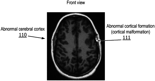
automatically detecting a cortical malformation based on the quantified scan data;
mapping a three-dimensional representation of the cerebral cortex to the quantified scan data to produce a mapped image of the cerebral cortex;
color-coding the mapped image of the cerebral cortex so that the cortical malformation is shown in a different color than a remainder of the cerebral cortex in the mapped image; and
outputting the color-coded mapped image of the cerebral cortex.
|