| CPC A61B 6/545 (2013.01) [A61B 6/027 (2013.01); A61B 6/032 (2013.01); A61B 6/0407 (2013.01); A61B 6/06 (2013.01); A61B 6/405 (2013.01); A61B 6/4085 (2013.01); A61B 6/4233 (2013.01); A61B 6/4435 (2013.01)] | 15 Claims |
|
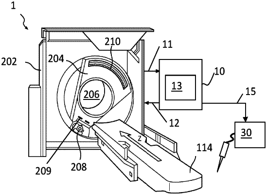
1. A device for controlling an image acquisition of a multi-slice computed tomography system comprising a rotatable gantry, a multi-row detector, an X-ray source and an automated examination couch moving in a longitudinal direction, the device comprising:
an input for receiving projection image data from the multi-row detector of the multi-slice computed tomography system;
an output for controlling operation of the multi-slice computed tomography system; and
a processor configured to control a first operation of the multi-slice computed tomography system via the output so that a computed tomography localizer radiograph of a subject to be imaged is acquired,
wherein the processor is adapted for receiving the computed tomography localizer radiograph via the input,
wherein the processor is adapted for controlling a second operation of the computed tomography system via the output so that a large volume overview that comprises at least an organ region defined on the computed tomography localizer radiograph and substantially delimiting an organ of interest in the subject is acquired, in which the subject is translated by the automated examination couch through an examination volume,
wherein controlling the second operation comprises increasing an X-ray cone angle of the computed tomography system in the longitudinal direction when the translation of the subject by the automated examination couch causes the organ region to enter into the examination volume and decreasing the X-ray cone angle when the translation of the subject causes the organ region to leave the examination volume.
|