| CPC C12Q 1/6886 (2013.01) [G01N 1/31 (2013.01); G01N 15/1475 (2013.01); G01N 33/5094 (2013.01); G01N 33/57484 (2013.01); G01N 33/582 (2013.01); C12Q 1/6883 (2013.01); G01N 15/1468 (2013.01); G01N 2015/1472 (2013.01); G01N 2333/4742 (2013.01); G01N 2333/70589 (2013.01); G06T 7/0012 (2013.01); G06T 2207/10056 (2013.01); G06T 2207/30096 (2013.01); G06T 2207/30101 (2013.01)] | 16 Claims |
|
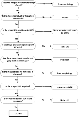
1. A computer program product for identifying candidate target cells within a biological fluid specimen, the computer program product tangibly embodied in a non-transitory computer readable medium, comprising instructions to cause a processor to
receive a digital image of the biological fluid specimen, the digital image having a plurality of color channels, wherein
the biological fluid specimen comprises a given cell in which a nucleus is completely contained within cytoplasm, and
the digital image comprises a representative of the given cell that is recorded with a visual artifact causing the digital image to represent the nucleus as partially contained within the cytoplasm and partially outside of the cytoplasm;
identify first connected regions of pixels of a minimum first intensity in a first channel of the plurality of color channels;
identify second connected regions of pixels of a minimum second intensity in a second channel of the plurality of color channels;
determine first connected regions and second connected regions that contain connected pixels indicating at least a partial spatial overlap; and
for each of least one of one or more pairs of a first connected region and a second connected region that contain connected pixels indicating at least a partial spatial overlap, account for the visual artifact comprising determining whether the second connected region overlaps the first connected region by a threshold amount, and if the second connected region overlaps the first connected region by the threshold amount then continue to treat a portion of the image corresponding to the overlap of first connected region and the second connected region as a candidate for classification.
|