| CPC B01L 3/502715 (2013.01) [A61B 5/157 (2013.01); A61B 5/150022 (2013.01); A61B 5/15113 (2013.01); A61B 5/15186 (2013.01); A61B 5/150213 (2013.01); A61B 5/150221 (2013.01); A61B 5/150274 (2013.01); A61B 5/150305 (2013.01); A61B 5/150343 (2013.01); A61B 5/150351 (2013.01); A61B 5/150412 (2013.01); A61B 5/150503 (2013.01); A61B 5/150786 (2013.01); B01L 3/5027 (2013.01); B01L 3/5029 (2013.01); B01L 3/5082 (2013.01); B01L 3/502738 (2013.01); B01L 3/502761 (2013.01); G01N 21/03 (2013.01); G01N 21/253 (2013.01); G01N 21/6452 (2013.01); G01N 21/6458 (2013.01); G01N 33/54373 (2013.01); G01N 35/0098 (2013.01); G01N 35/02 (2013.01); G01N 35/025 (2013.01); A61B 5/117 (2013.01); A61B 5/151 (2013.01); B01L 7/00 (2013.01); B01L 9/52 (2013.01); B01L 2200/025 (2013.01); B01L 2200/026 (2013.01); B01L 2200/027 (2013.01); B01L 2200/0605 (2013.01); B01L 2200/10 (2013.01); B01L 2300/022 (2013.01); B01L 2300/042 (2013.01); B01L 2300/043 (2013.01); B01L 2300/046 (2013.01); B01L 2300/048 (2013.01); B01L 2300/0654 (2013.01); B01L 2300/0681 (2013.01); B01L 2300/0864 (2013.01); B01L 2400/043 (2013.01); B01L 2400/0406 (2013.01); B01L 2400/0409 (2013.01); B01L 2400/0415 (2013.01); B01L 2400/0457 (2013.01); B01L 2400/0472 (2013.01); B01L 2400/0481 (2013.01); B01L 2400/0677 (2013.01); B01L 2400/0683 (2013.01); G01N 21/6428 (2013.01); G01N 2021/0325 (2013.01); G01N 2021/0346 (2013.01); G01N 2035/0401 (2013.01); G01N 2035/0429 (2013.01)] | 27 Claims |

|
1. An automated imaging analyzer for detecting individual microscopic targets at low magnification, the automated image analyzer comprising:
a sample cartridge, the sample cartridge comprising a sample input reservoir having a sample therein, a first plurality of wells configured for fluid communication with the sample input reservoir, and a plurality of imaging wells, wherein each imaging well of the plurality of imaging wells is configured for fluid communication with a respective well of the first plurality of wells, each imaging well comprising a detection surface;
a housing configured to accept the sample cartridge;
a reagent processing subsystem operable to:
mobilize the sample within the sample input reservoir of the sample cartridge to disperse the sample into the first plurality of wells through a first plurality of channels of the sample cartridge, and
mobilize the sample from each well of the first plurality of wells of the sample cartridge into a respective imaging well of the plurality of imaging wells of the sample cartridge through a second plurality of channels of the cartridge,
wherein the reagent processing subsystem is configured to actuate at least one valve of the sample cartridge and apply a pressure gradient to the sample cartridge to cause flow of the sample within the sample cartridge to disperse the sample into the first plurality of wells, and
a magnetic station comprising a magnet connected to the housing and disposed to apply a selective force to move magnetic particles in each respective imaging well to deposit the magnetic particles on said detection surface of the respective imaging well;
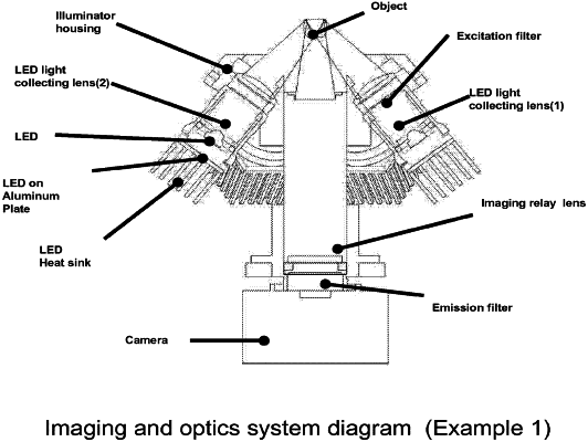
an imaging station comprising a collecting lens arranged to collect optical signal from the detection surface;
a photoelectric array detector connected to the housing and disposed to receive the optical signal and produce an image of individual particles deposited on the detection surface; and
a conveyor operable to move the sample cartridge between a plurality stations within the housing, the plurality of stations comprising the imaging station and the magnetic station,
wherein the image has lower than 5-fold magnification, and wherein the individual microscopic targets are detectable within the image.
|