| CPC A61M 27/006 (2013.01) [B33Y 80/00 (2014.12); A61M 2205/0216 (2013.01); A61M 2205/0294 (2013.01); A61M 2205/3306 (2013.01); A61M 2205/3313 (2013.01); A61M 2205/3334 (2013.01); A61M 2205/702 (2013.01); A61M 2207/00 (2013.01); A61M 2210/0693 (2013.01)] | 21 Claims |
|
1. A system, comprising:
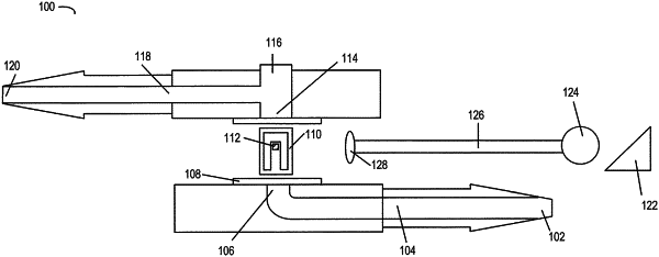
a fluidic device adapted to be positioned under a skin of a patient, the fluidic device comprising:
a first channel including a first inlet and a first outlet;
a second channel including a second inlet and a second outlet, wherein the second inlet of the second channel is in fluid communication with the first outlet of the first channel;
a sensor positioned between the first outlet and the second inlet, wherein the sensor comprises a membrane valve configured to deflect from a first position to a second position in response to a flow between the first channel and the second channel, and wherein the membrane valve includes one or more biocompatible reflective surfaces; and
an optical system adapted to be positioned under the skin of the patient, wherein the optical system includes a focusing lens configured to focus light from a light source onto the membrane valve, wherein the one or more biocompatible reflective surfaces of the membrane valve are configured to deflect light from the membrane valve to an imaging device, wherein the imaging device is configured to measure a light intensity of light reflected off of the one or more biocompatible reflective surfaces of the membrane valve, and wherein the light intensity measured by the imaging device has a first value when the membrane valve is in the first position, wherein the light intensity measured by the imaging device has a second value when the membrane valve is in the second position in response to the flow between the first channel and the second channel, and wherein the second value is greater than or less than the first value.
|