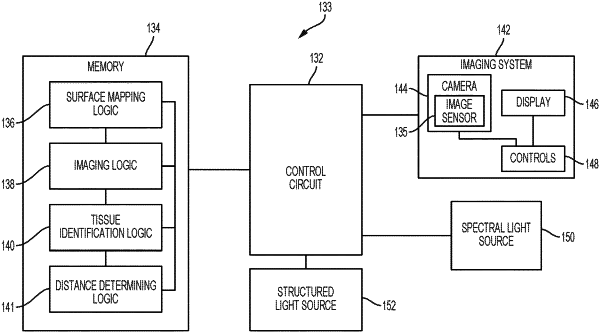
| CPC A61B 90/361 (2016.02) [A61B 1/009 (2022.02); A61B 1/000094 (2022.02); A61B 1/00194 (2022.02); A61B 1/046 (2022.02); A61B 1/0605 (2022.02); A61B 34/10 (2016.02); G06T 17/00 (2013.01); A61B 2034/105 (2016.02); A61B 2034/107 (2016.02); G06T 2200/04 (2013.01)] | 18 Claims |

|
1. A surgical visualization system comprising:
a plurality of light sources;
at least one optical sensor;
at least one display device; and
a controller comprising a processor and a memory unit, wherein the memory unit is configured to store instructions that, when executed by the processer, cause the controller to:
control at least one of the plurality of light sources to illuminate a tissue in a first manner by projecting a pattern of light onto a surface of the tissue;
control the at least one optical sensor to receive first light reflected by the surface of the tissue when illuminated in the first manner;
calculate first imaging data of a three-dimensional image of one or more surface features of the tissue based on the first light reflected by the surface of the tissue when illuminated in the first manner;
control at least one of the plurality of light sources to illuminate the tissue in a second manner by illuminating the tissue with a plurality of emitted light waves, wherein each of the plurality of emitted light waves has a unique light spectrum, and wherein at least one of the plurality of emitted light waves has a light spectrum configured based on a spectral signature of one or more subsurface critical anatomic structures of the tissue;
control the at least one optical sensor to receive second light from the tissue when illuminated in the second manner;
calculate second imaging data of the one or more subsurface critical anatomic structures of the tissue based on the second light received by the at least one optical sensor; and
display a combination of the first imaging data and the second imaging data on the at least one display device.
|