| CPC A61B 1/000094 (2022.02) [A61B 1/009 (2022.02); A61B 1/00194 (2022.02); A61B 1/046 (2022.02); A61B 1/0605 (2022.02); A61B 90/361 (2016.02); A61B 90/37 (2016.02); G06T 7/30 (2017.01); G06T 7/521 (2017.01); G06T 19/00 (2013.01); A61B 17/064 (2013.01); A61B 34/25 (2016.02); A61B 34/30 (2016.02); A61B 2090/064 (2016.02); A61B 2090/367 (2016.02); A61B 2090/3735 (2016.02); G06T 2200/24 (2013.01); G06T 2207/10081 (2013.01); G06T 2207/10088 (2013.01); G06T 2207/20221 (2013.01); G06T 2207/30092 (2013.01); G06T 2210/41 (2013.01); G06T 2219/004 (2013.01)] | 26 Claims |
|
1. A surgical visualization system, comprising:
a display screen;
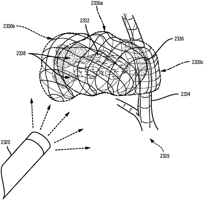
a first source configured to emit a plurality of spectral light waves configured to penetrate an organ;
a second source configured to emit a structured light pattern onto a surface of the organ;
an image sensor comprising a three-dimensional camera, wherein the image sensor is configured to detect at least one of the plurality of spectral light waves that are reflected or the structured light pattern that is reflected; and
a control circuit in signal communication with the first source, the second source, the image sensor, and the display screen, wherein the control circuit is configured to:
receive pre-operative imaging data associated with the organ;
receive first intraoperative imaging data indicative of the structured light pattern on the surface of the organ;
generate a three-dimensional digital representation of the organ based on the pre-operative imaging data and the first intraoperative imaging data;
display the three-dimensional digital representation of the organ on the display screen;
receive second intraoperative imaging data indicative of the plurality of spectral light waves penetrating the organ;
intraoperatively identify a structure below the surface of the organ based on the second intraoperative imaging data;
generate a three-dimensional digital representation of the structure below the surface of the organ; and
overlay the three-dimensional digital representation of the structure with the three-dimensional digital representation of the organ on the display screen.
|