| CPC G06T 7/0014 (2013.01) [G06T 7/11 (2017.01); G06T 7/30 (2017.01); G06T 2207/10081 (2013.01); G06T 2207/20081 (2013.01); G06T 2207/20084 (2013.01); G06T 2207/20212 (2013.01); G06T 2207/30016 (2013.01)] | 20 Claims |

|
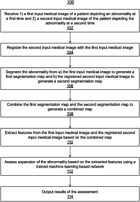
1. A computer-implemented method comprising:
receiving 1) a first input medical image of a patient depicting an abnormality at a first time and 2) a second input medical image of the patient depicting the abnormality at a second time;
registering the second input medical image with the first input medical image;
segmenting the abnormality from a) the first input medical image to generate a first segmentation map and b) the registered second input medical image to generate a second segmentation map;
combining the first segmentation map and the second segmentation map to generate a combined map;
extracting features from the first input medical image and the registered second input medical image based on regions in which the abnormality is located, wherein the regions are identified by the combined map;
assessing expansion of the abnormality based on the extracted features using a trained machine learning based network; and
outputting results of the assessment.
|