| CPC A61B 8/485 (2013.01) [A61B 8/085 (2013.01); A61B 8/4494 (2013.01); B06B 1/0622 (2013.01); G01S 7/52038 (2013.01); G01S 15/8913 (2013.01); G01S 15/8922 (2013.01); G01S 15/8952 (2013.01); A61B 8/5207 (2013.01); G01S 7/5203 (2013.01)] | 14 Claims |
|
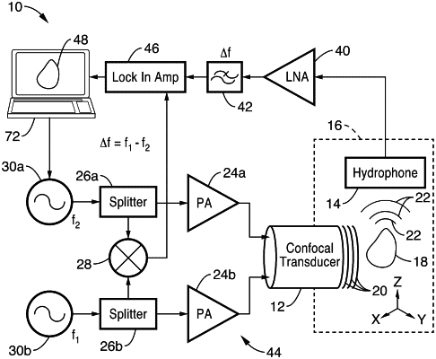
1. A method for performing multi-frequency harmonic acoustography for target identification and border detection within a target tissue, the method comprising:
providing a focused confocal transducer having at least one piezoelectric element;
focusing first and second ultrasonic waves at first and second frequencies from the transducer on the target tissue;
wherein the first and second waves interfere at a focal plane within the target tissue such that the target tissue absorbs energy from the first and second waves and vibrates to emit a third acoustic wave within the target tissue;
detecting the third acoustic wave with a hydrophone; and
analyzing the third acoustic wave to evaluate one or more mechanical properties of the target tissue by analyzing harmonics generated by non-linear properties of the target tissue, wherein a spectral envelope comprising relative strengths of higher harmonics is used to create imaging contrast and unique identifying information associated with the target tissue; and
wherein the one or more mechanical properties of the target tissue are selected from the group consisting of: tissue type, size, location with adjacent tissue or physiologic or disease state.
|