| CPC A61B 5/0095 (2013.01) [A61B 1/041 (2013.01); A61B 1/00147 (2013.01); A61B 1/00148 (2022.02); A61B 1/00165 (2013.01); A61B 5/0066 (2013.01); A61B 5/0068 (2013.01); A61B 5/0071 (2013.01); A61B 5/0075 (2013.01); A61B 5/6861 (2013.01); A61B 8/12 (2013.01)] | 14 Claims |

|
1. A device for endoscopic optoacoustic imaging, the device comprising:
an imaging unit configured to be at least partially inserted into an object, the imaging unit comprising:
an irradiation unit configured to irradiate a region of interest inside the object with electromagnetic radiation, and
a detection unit comprising at least one ultrasound transducer configured to detect ultrasound waves generated in the region of interest in response to irradiating the region of interest with the electromagnetic radiation and to generate detection signals, the at least one ultrasound transducer exhibiting a field of view being at least partially located in the irradiated region of interest,
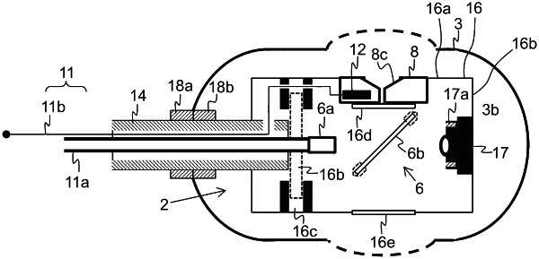
a position stabilizing structure forming a housing for at least a part of the imaging unit, the position stabilizing structure comprising an outer face and an interior and being configured to stabilize and/or fix the imaging unit in a position and/or orientation in the object by bringing the outer face of the position stabilizing structure into contact with the object,
a processing unit configured to generate an optoacoustic image of the region of interest based on the detection signals; and
a carrier unit disposed in the interior of the position stabilizing structure, the carrier unit comprising a proximal end, a distal end, a first lateral face, and a second lateral face, wherein the irradiation unit and the detection unit are mounted on the carrier unit such that the electromagnetic radiation emanating from the irradiation unit is directed towards the field of view of the at least one ultrasound transducer, the detection unit being mounted on the first lateral face and being oriented substantially perpendicular to a longitudinal axis of the position stabilizing unit, the at least one ultrasound transducer exhibiting a sensitive surface, wherein the at least one ultrasound transducer is mounted and/or arranged on the carrier unit such that the sensitive surface of the ultrasound transducer faces towards the region of interest inside the object, wherein the at least one at least one ultrasound transducer comprises an aperture or window section being at least partially transparent to the electromagnetic radiation; and
an optical fiber and a reflection element, wherein a distal end of the optical fiber is disposed within the carrier unit between the proximal end of the carrier unit and the reflection element, wherein the reflection element is arranged within the carrier unit between the distal end of the optical fiber and the ultrasound transducer and is configured to reflect the electromagnetic radiation emanating from the distal end of the optical fiber such that the reflected electromagnetic radiation passes through the aperture or window section of the ultrasound transducer to irradiate the region of interest inside the object.
|