| CPC A61B 5/0044 (2013.01) [A61B 5/02028 (2013.01); A61B 5/7267 (2013.01); A61B 5/7278 (2013.01); G01R 33/56 (2013.01); G06N 3/08 (2013.01); G06T 7/0012 (2013.01); G06T 7/11 (2017.01); G16H 30/40 (2018.01); A61B 5/055 (2013.01); A61B 2576/023 (2013.01); G06T 2207/10088 (2013.01); G06T 2207/20081 (2013.01); G06T 2207/20084 (2013.01); G06T 2207/30048 (2013.01)] | 17 Claims |

|
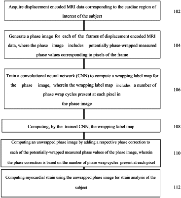
1. A method of strain analysis of a cardiac region of interest of a subject from displacement encoded magnetic resonance image (MRI) data, the method comprising:
acquiring displacement encoded MRI data corresponding to the cardiac region of interest of the subject;
generating a phase image for each frame of the displacement encoded MRI data, wherein the phase image comprises potentially phase-wrapped measured phase values corresponding to pixels of the frame;
training a convolutional neural network (CNN) to compute a wrapping label map for the phase image, wherein the wrapping label map comprises a number of phase wrap cycles present at each pixel in the phase image;
computing, by the trained CNN, the wrapping label map;
computing an unwrapped phase image by adding a respective phase correction to each of the potentially phase-wrapped measured phase values of the phase image, wherein the phase correction is based on the number of phase wrap cycles present at each pixel; and
computing myocardial strain using the unwrapped phase image for strain analysis of the subject.
|