| CPC A61B 6/481 (2013.01) [A61B 6/032 (2013.01); A61B 6/4241 (2013.01); A61B 6/469 (2013.01); A61B 6/50 (2013.01); A61B 6/5205 (2013.01); A61M 5/007 (2013.01)] | 19 Claims |
|
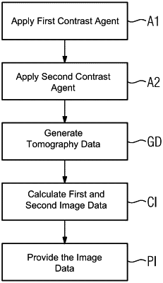
1. A method for providing image data of a hollow organ of a digestive system, comprising:
applying a first contrast agent to a lumen of the hollow organ to obtain a contrast agent filling of the lumen, the first contrast agent having a first absorption spectrum;
applying a second contrast agent to a blood vessel system supplying a wall surrounding the lumen of the hollow organ, the second contrast agent providing substantially complete perfusion of the wall, and the second contrast agent having a second absorption spectrum different from the first absorption spectrum;
generating spectrally resolved computed tomography data of an examination area including the hollow organ;
calculating first image data and second image data by applying a material separation algorithm onto the spectrally resolved computed tomography data, the first image data indicative of a presence of the first contrast agent, and the second image data indicative of a presence of the second contrast agent;
generating a representation of the wall based on the first image data and the second image data, the generating the representation of the wall including assigning a pixel of the second image data to the representation of the wall based on a distance of the pixel from a representation of a border of the first contrast agent filling of the lumen; and
providing the representation of the wall.
|